
Best Diagnostic Centre in KumbakonamAdvanced CT Scan & Ultrasound Diagnostic Centre

-
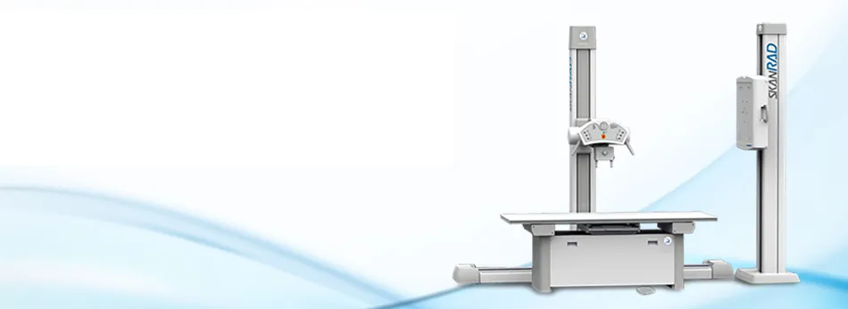
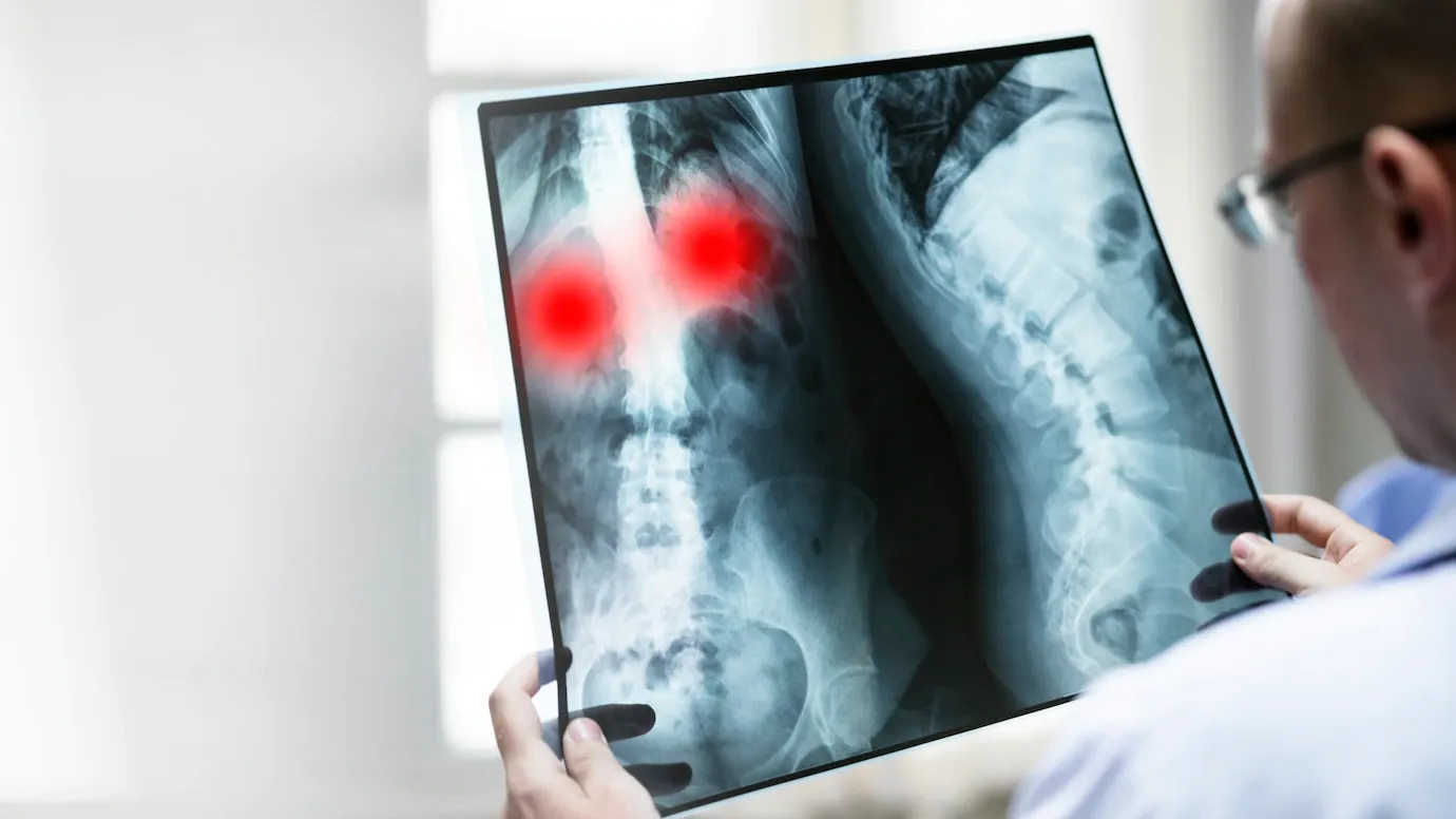
X-Ray
X-rays use a type of radiation called electromagnetic waves to produce images of structures in our body like joints and bones.
-
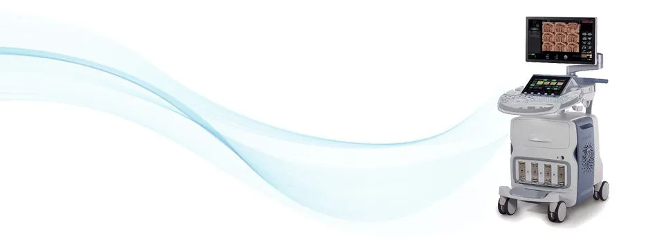
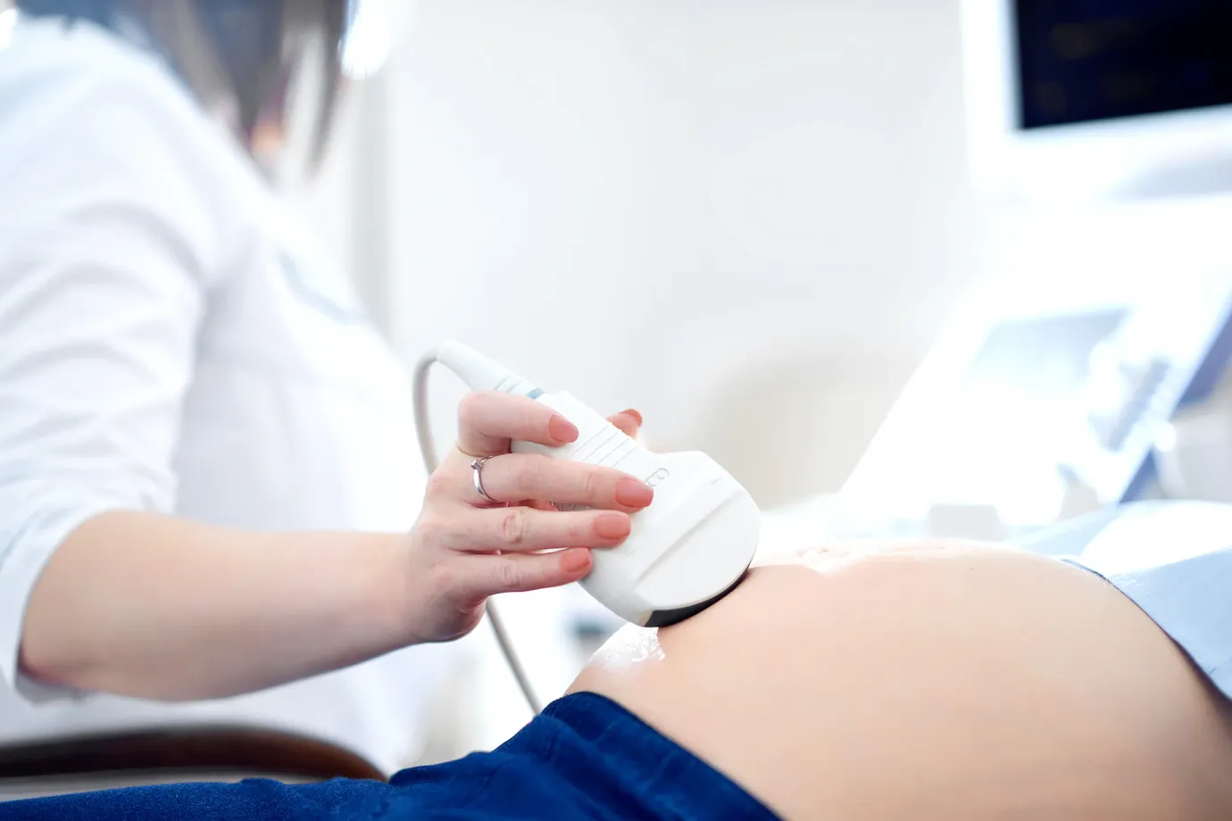
Ultrasound
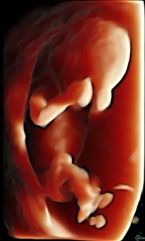
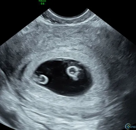
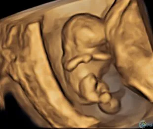
Ultrasound scanning is a clinical tool which provides images of internal fetal anatomy, as well as a wide range of other organ systems.
-
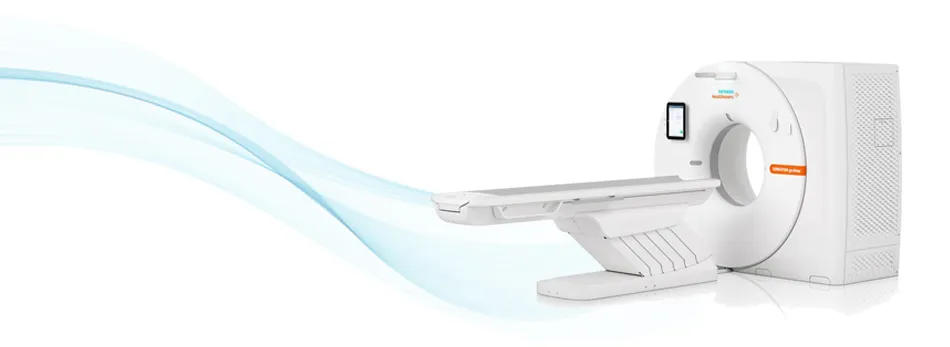
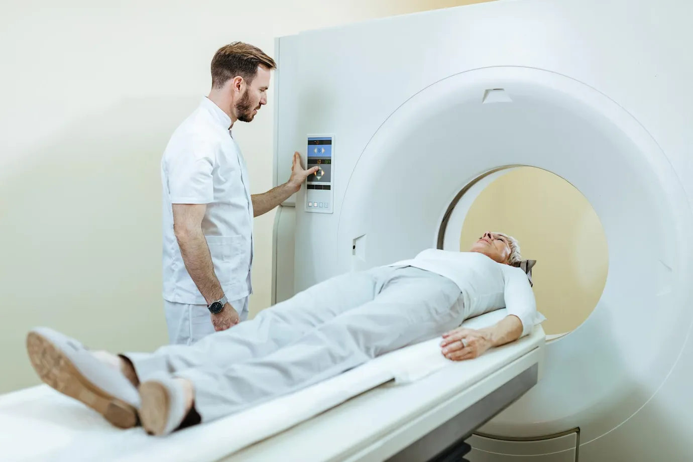
CT Scan & CT Angiogram
CT scan & angio can show detailed images of any part of the body, bones, muscles, organs and blood vessels.
A Modern, Computerized Scan Centre
Dr. Kalirajan Diagnostics is a well-recognized scan centre for its best radiological services, located in Kumbakonam. We serve for the betterment of patients' health by providing accurate reports and test packages at an affordable price.
Computerised scan centre
Affordable price
365 days service

Tests by Condition
Different body part X-rays and scans are available and the results are shown accurately.
- Thyroid
- Liver
- Lungs
- Infertility
- Kidney
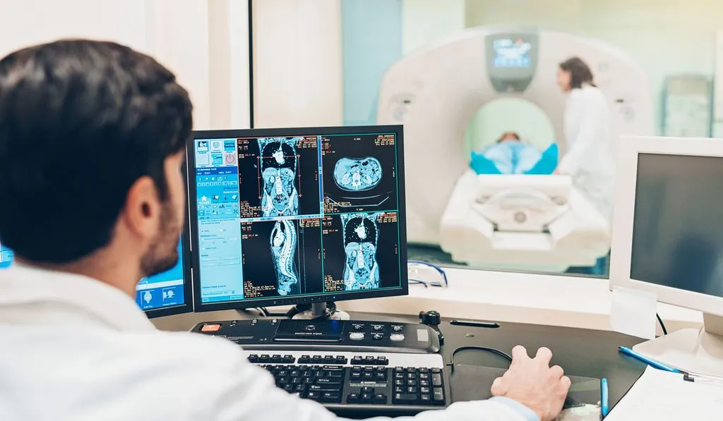
Our Expertise
To deliver the most accurate diagnosis.
-
X-Ray
- All body part X-Ray
- Barium special investigations
-
Ultrasound
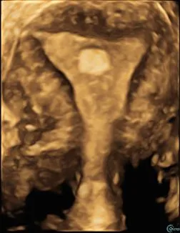
- Abdomen & Pelvis
- Small Parts
- Obstetrics Scan
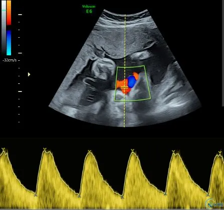
- Doppler Studies
-
CT Scan
- Brain, PNS, Orbits, HRCT temporal bone, Facial bones, Neck
- HRCT chest, Abdomen and pelvis, KUB
- Pelvic bones, Spine, Joints, Long bones
- Region specific, Fistulogram
-
CT Angiogram
- Neck and cerebral, Aortogram
- Pulmonary, Renal and Abdomen angiogram
- Liver angiogram
- Peripheral angiogram (upper / lower limb)
Get in Touch
Place your queries and doubts
- Call90433 72380
- Call0435 4066047
- Address232, Kamaraj Road, Anna Nagar, Kumbakonam - 612001, Tamil Nadu
- Opening hoursMon-Sar: 9 am–8 pmSun: 9:30 am–12 pm
Send us your query
KRS Image Library
Typical, rare and difficult cases, radiological signs and spotters from our centre.
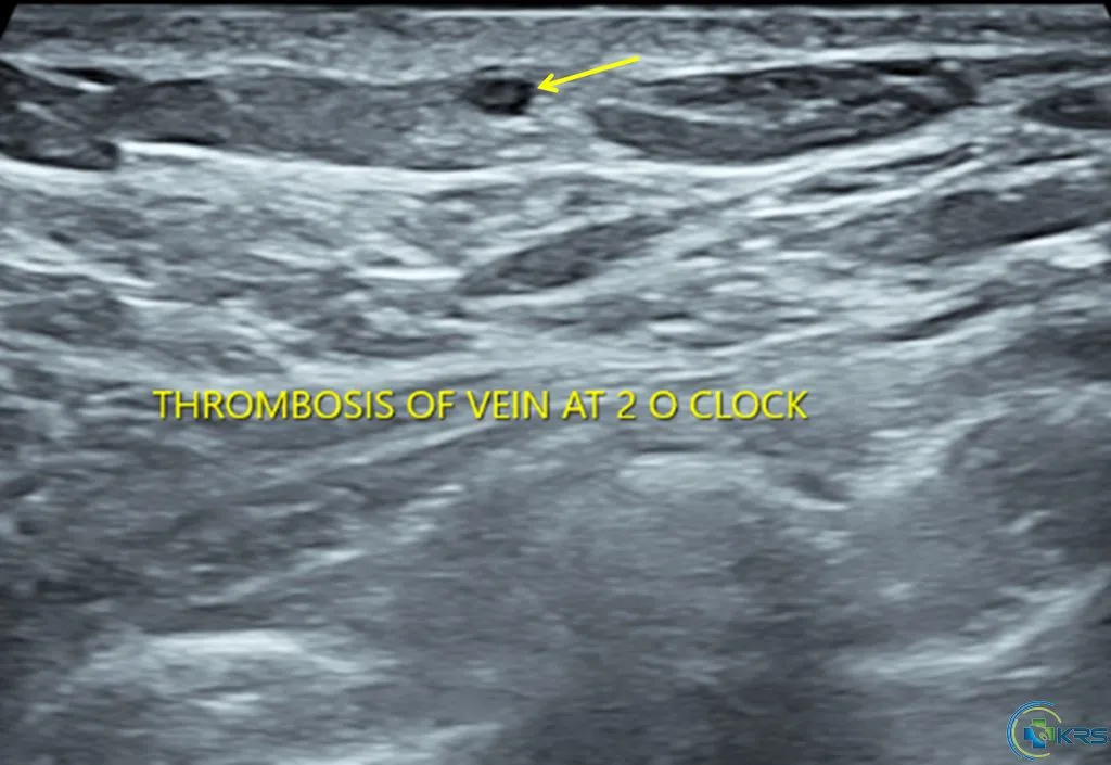
Mondor's disease
27 yr female presented with short duration of left breast pain.
- Focal thickening and echogenic lumen of superficial vein of left breast at 2 O'clock position with loss of compressibility (arrow) – suggestive of acute thrombophlebitis of superficial vein of breast – Mondor's disease.
- Rest of the breast reveal no focal lesion or mass lesion.
- No axillary lymphadenopathy.
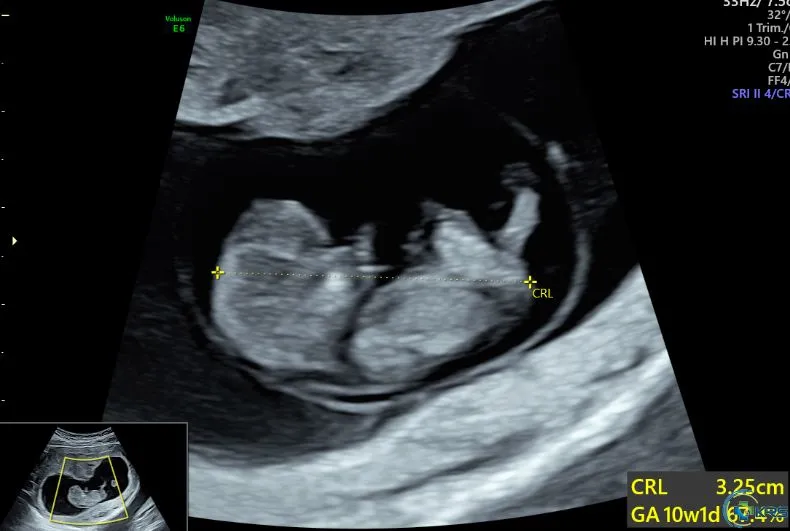
Physiological midgut herniation
20 yr female presented for routine dating scan at 10 weeks of pregnancy.
- Single live intrauterine fetus with CRL of 32.5 mm, corresponding to 10 weeks 1 day.
- Examination of the fetus revealed echogenic midgut herniation through umbilical defect (arrow) suggestive of physiological midgut herniation.
- Physiological midgut herniation – normally seen in fetus of 9 to 11 weeks. Abnormal if seen after 13 weeks of pregnancy.
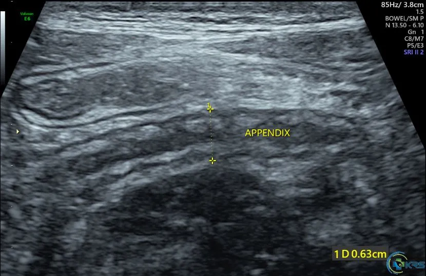
Acute appendicitis
18 yr male presented with acute abdominal pain for 2 days duration.
- Appendix – retroileal in location at 5 O'clock position, directing towards pelvis.
- Appendix is distended measuring 7.8 mm in maximum diameter and shows wall thickening.
- Periappendiceal inflammatory fat stranding (arrow) noted.
- No evidence of collection / abscess.
- Features consistent with acute appendicitis (uncomplicated).
Sample Images from KRS Image Library
Videos
From our YouTube channel.
Request an appointment
Choose a convenient date and time for your scan. Our team will call you to confirm.