10 yr boy presented with acute onset abdominal pain and vomiting.
Ultrasound findings:
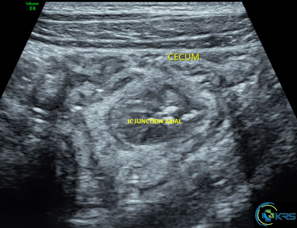
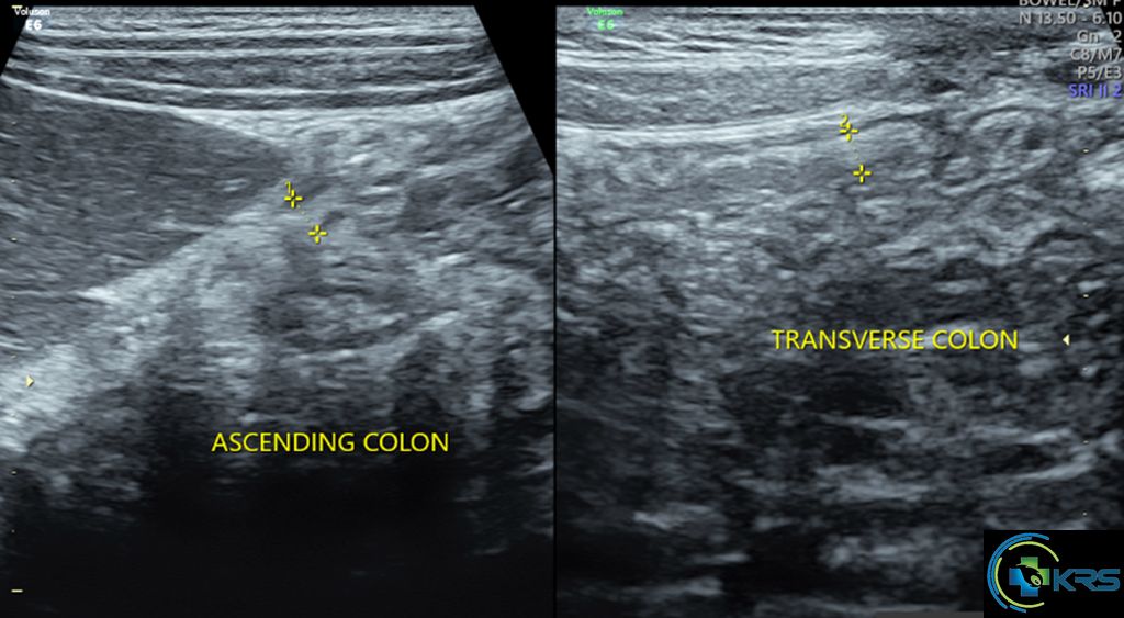
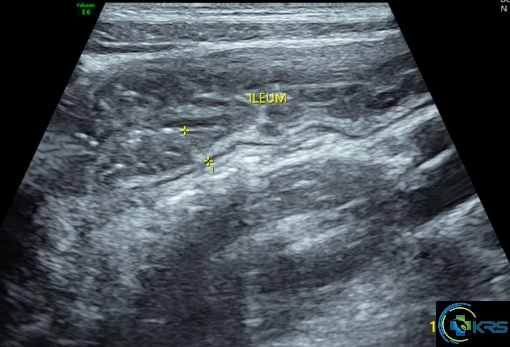
• Long segment circumferential wall thickening involving terminal ileum, cecum, ascending colon and transverse colon with maximum single wall thickness of 5.1 mm.
• No evidence of wall thickening of descending or sigmoid colon.
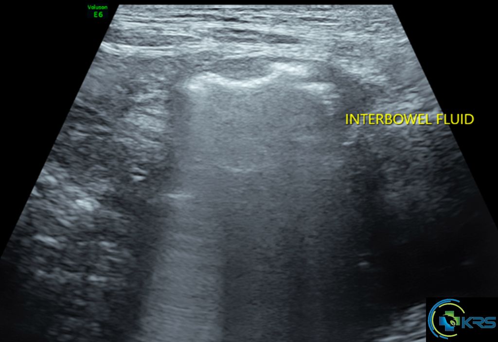
• Minimal interbowel fluid in pelvis.
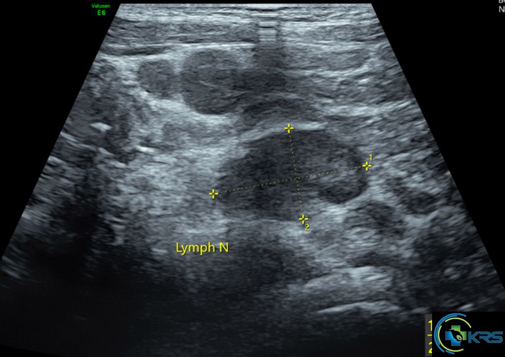
• Multiple enlarged ileo-colic lymphnodes, largest measuring 2.3 x 1.4 cm. No evidence of necrosis.
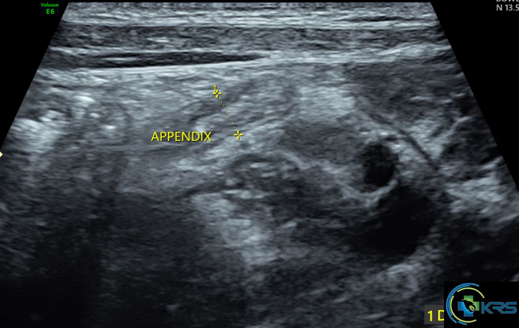
• Appendix measures 4.8 mm and appear normal.
• – Features suggestive of Acute ileo-colitis (enteritis).