This is a knowledge sharing segment intended to publish our typical/ rare/ difficult cases, radiological signs and spotters. Please feel free to contact in case of queries
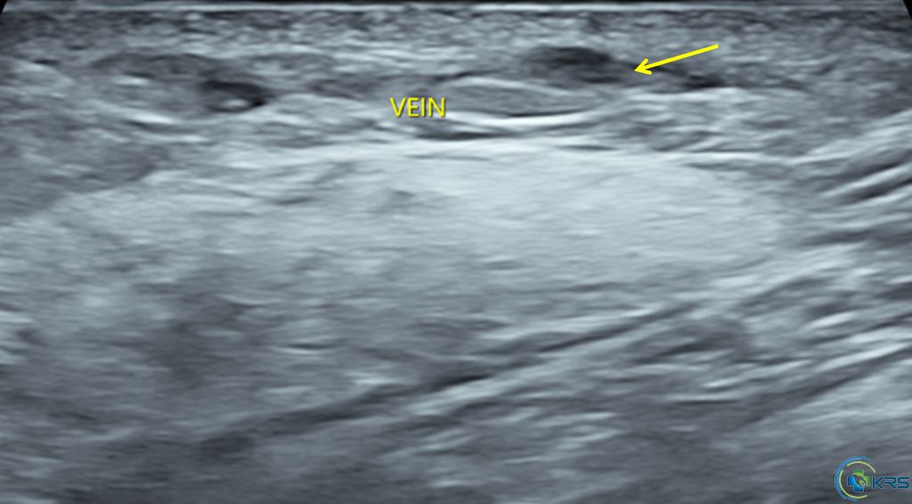
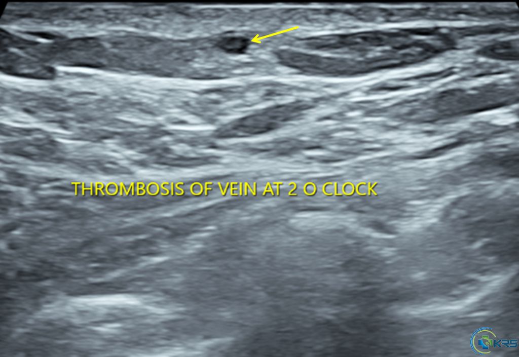
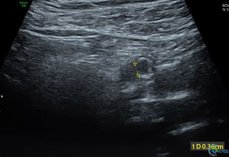
27 yr female presented with short duration of left breast pain.
Sonomammogram was done and revealed:
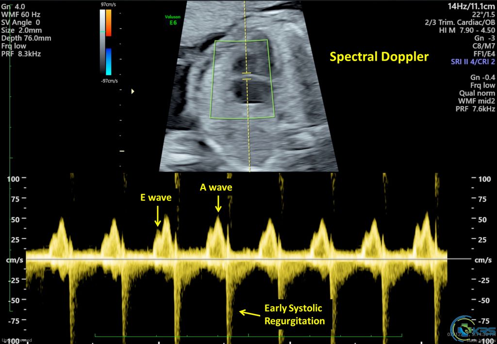
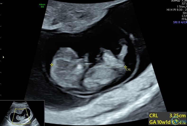
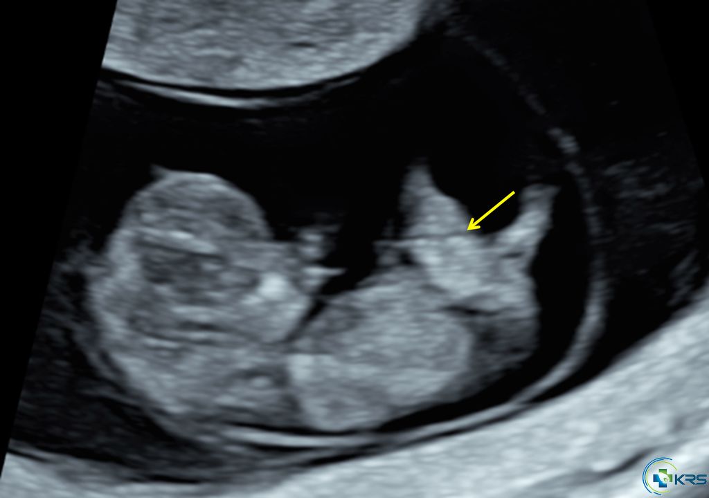
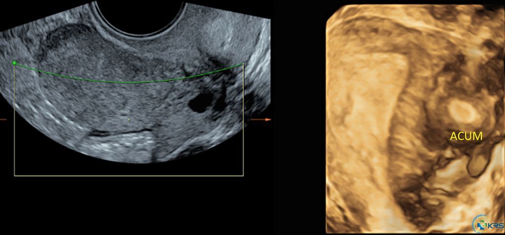
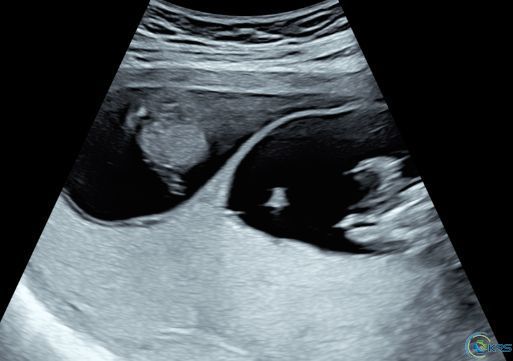
20 yr female presented for routine dating scan at 10 weeks of pregnancy
Ultrasound findings:
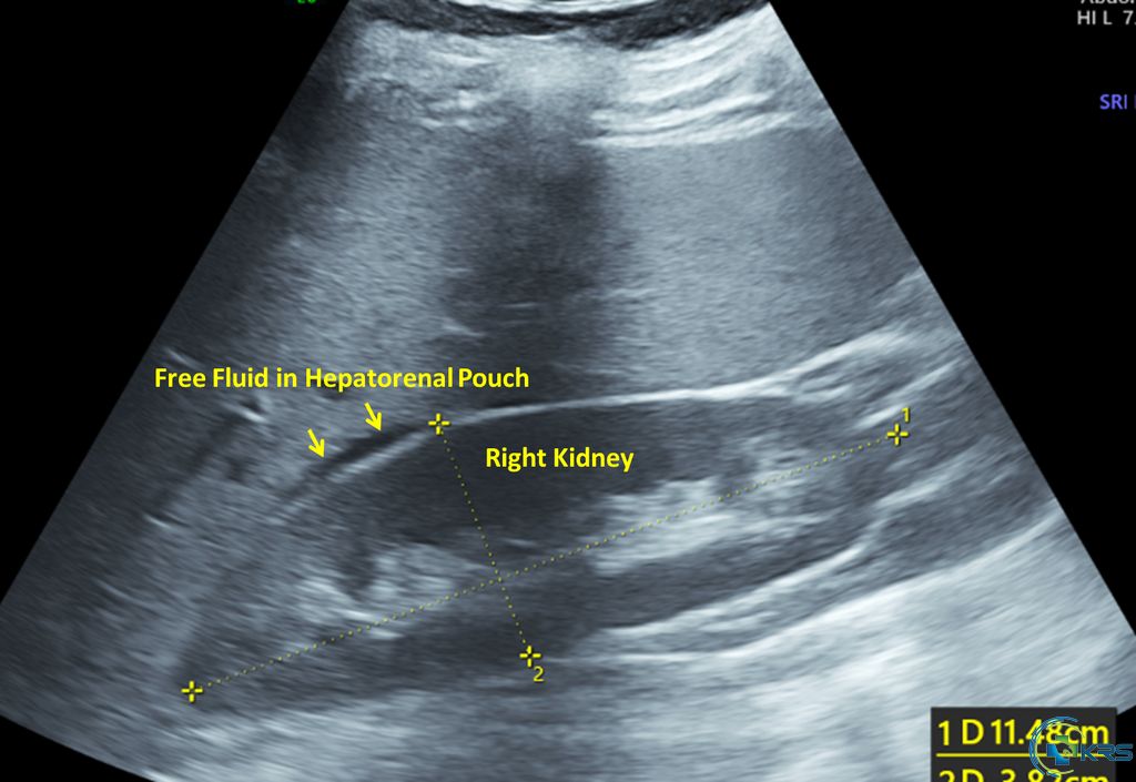
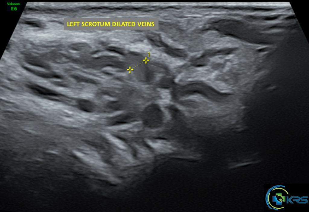
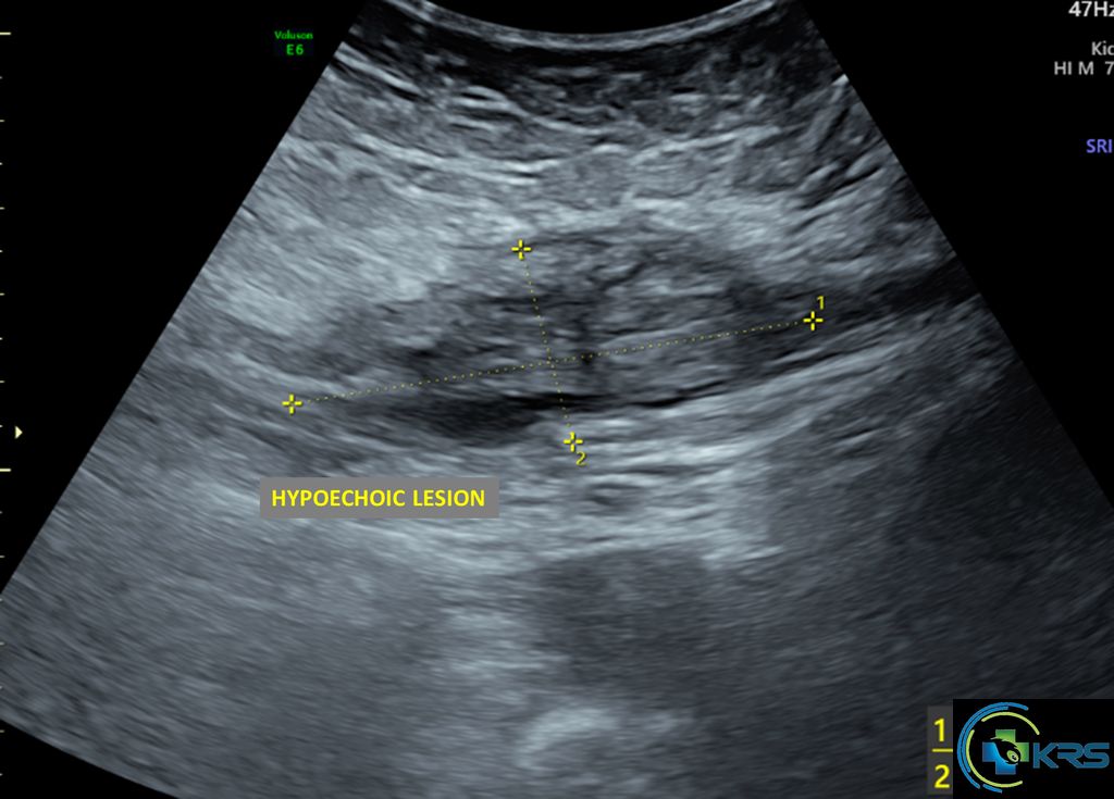
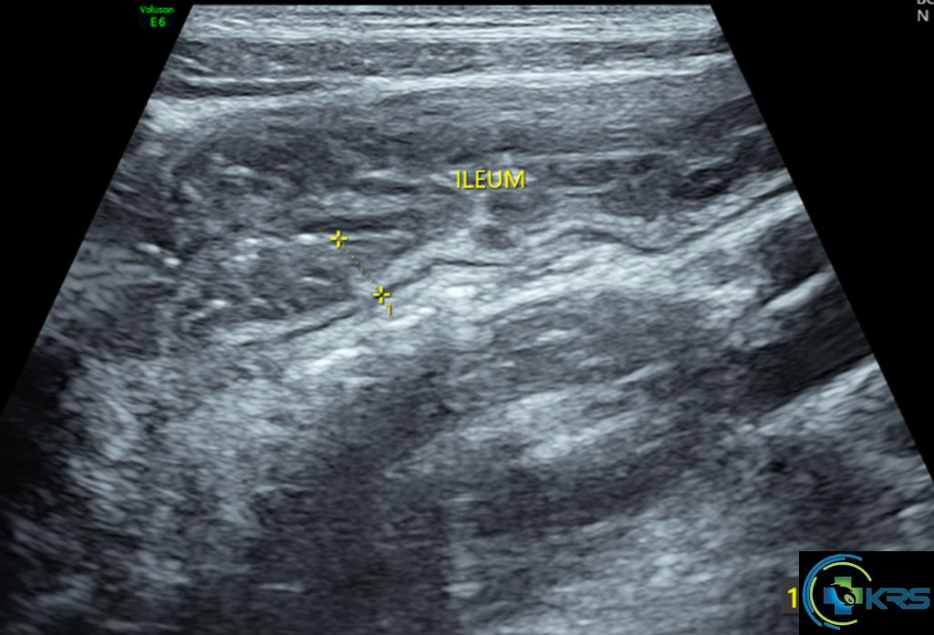
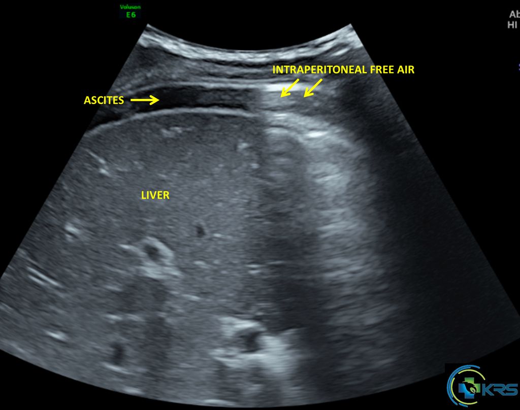
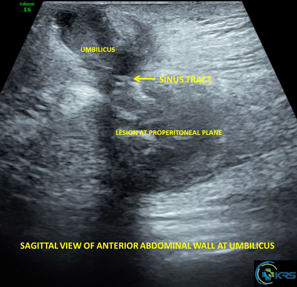
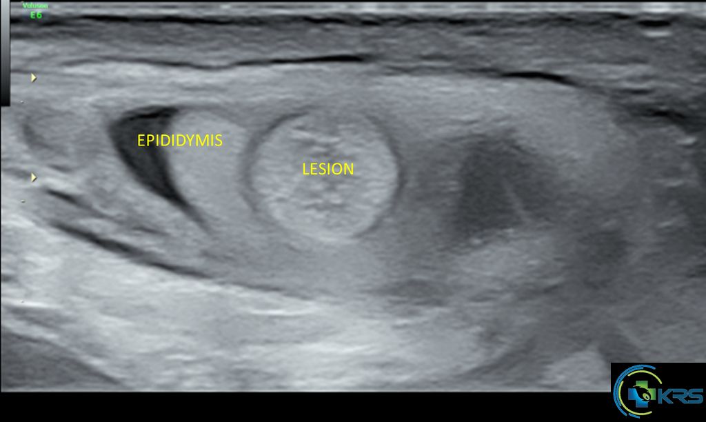
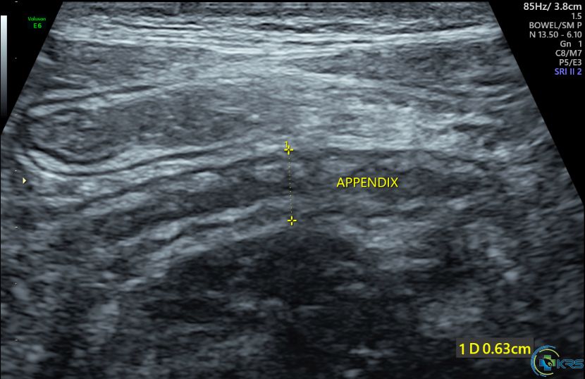
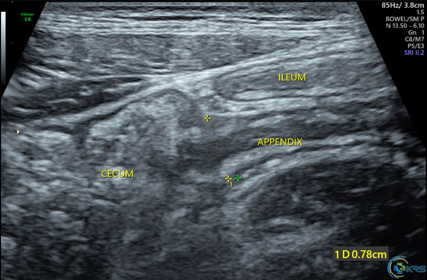
18 yr male presented with acute abdominal pain for 2 days duration
Ultrasound findings:
63 yr female presented with lower abdomen pain. Ultrasound abdomen showed aneurysm of abdominal aorta.
CECT abdomen findings:
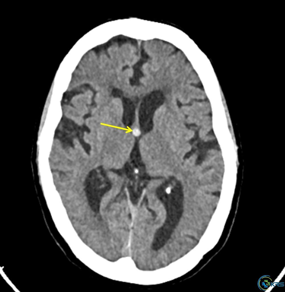
61 yr female came for routine screening
Ultrasound findings:
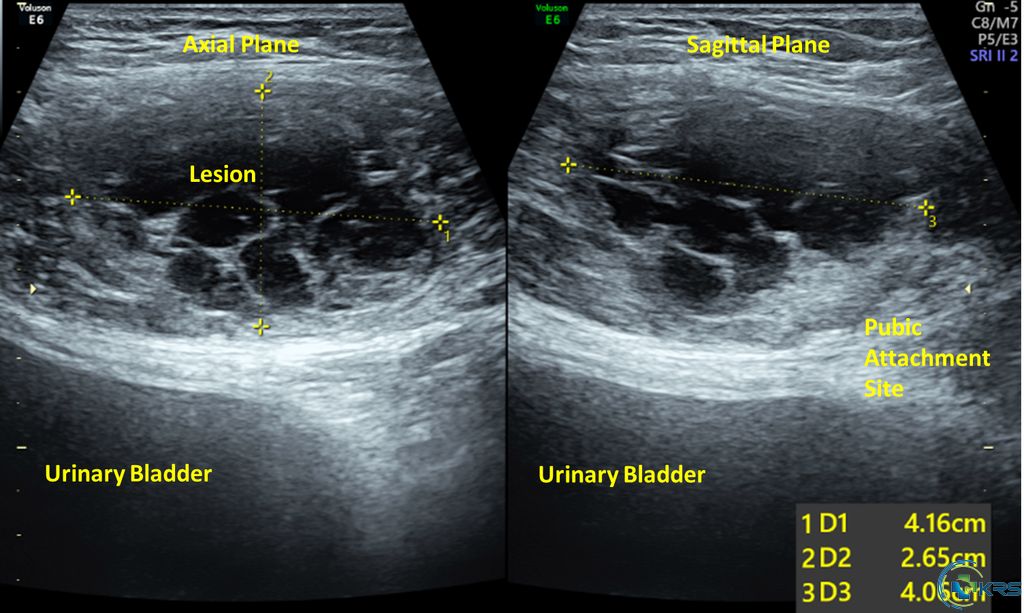
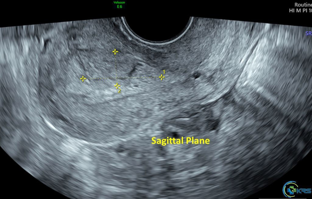
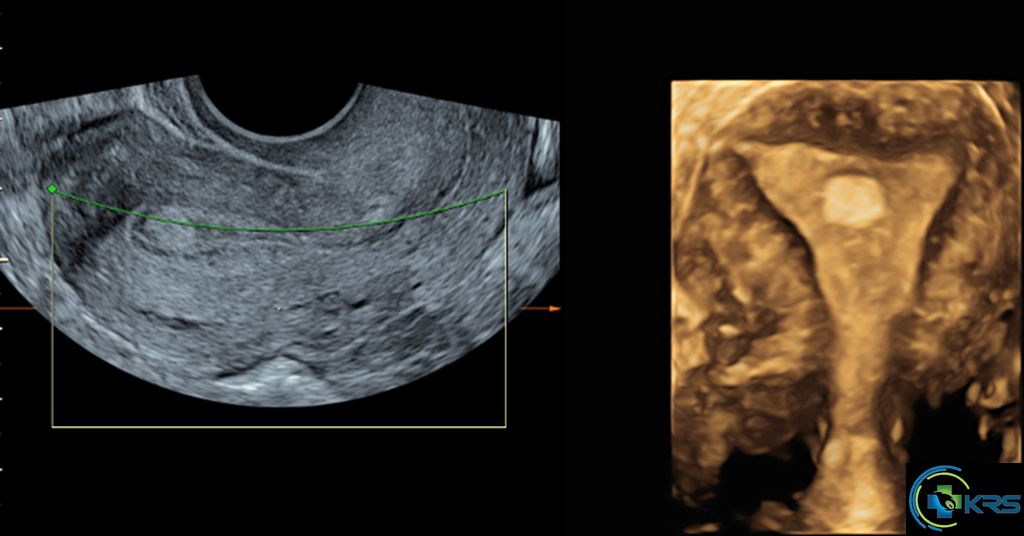
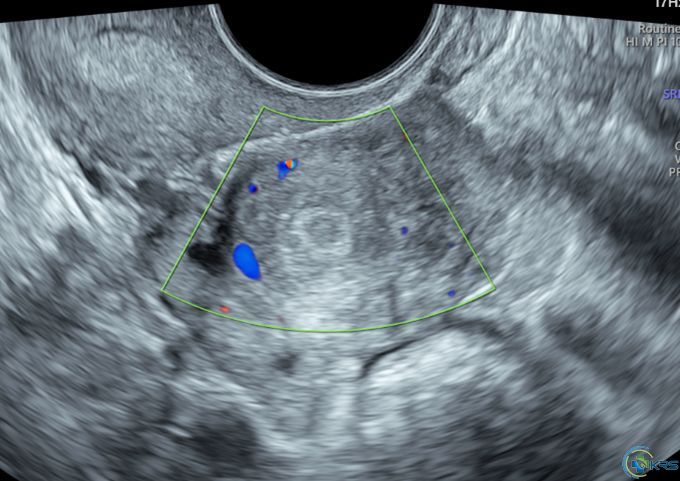
25 yr female with history of dysmenorrhea and infertility
Ultrasound findings:
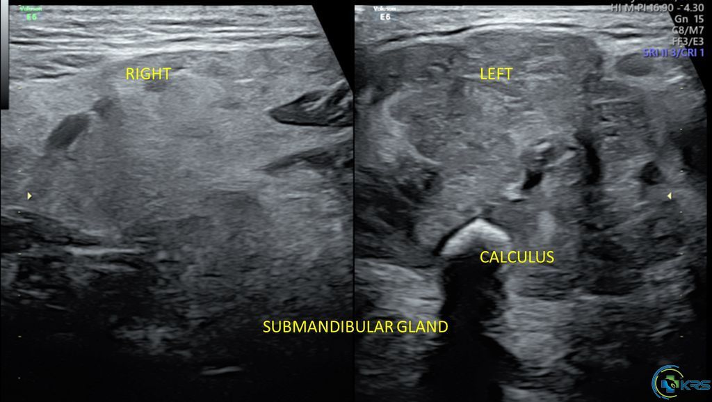
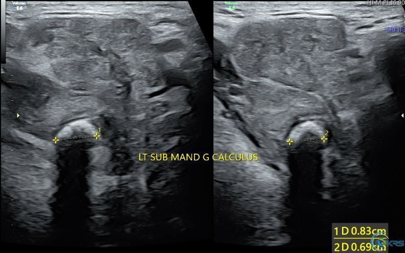
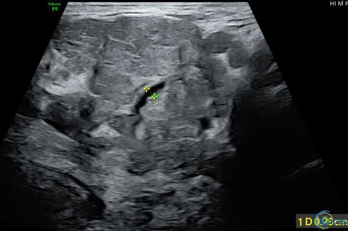
27 yr male complaining of swelling and pain in left submandibular region aggravated while eating
Ultrasound findings:
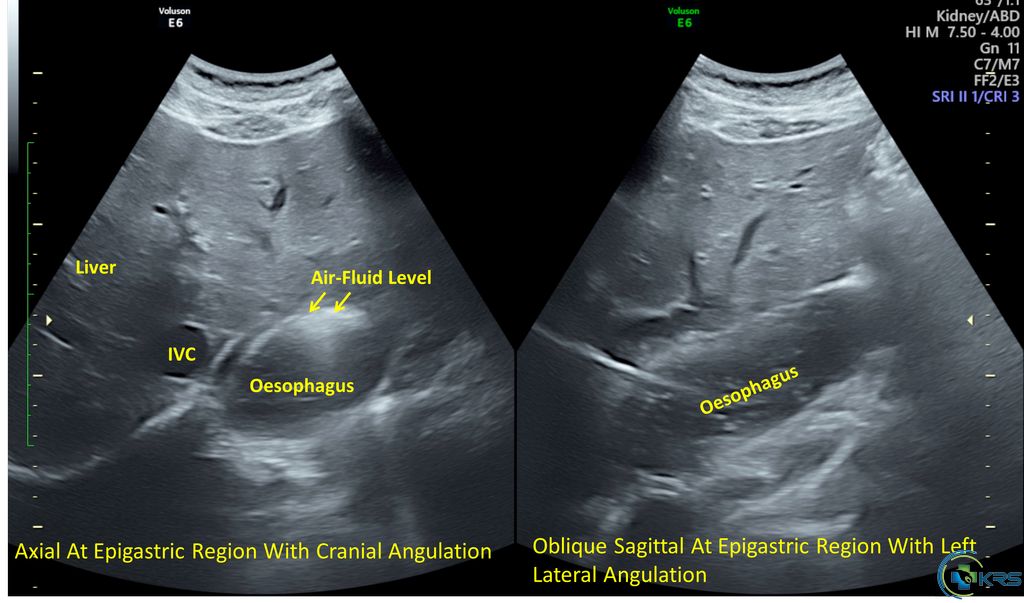
47 yr male presented with acute vomiting and abdominal pain
Ultrasound findings:
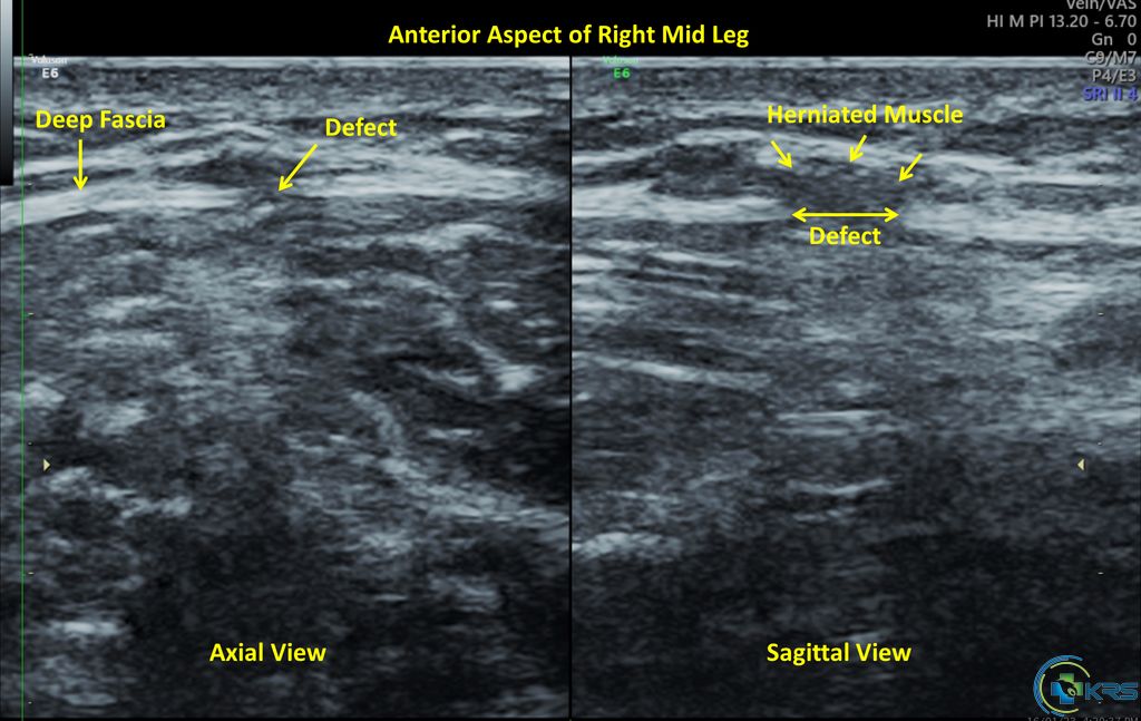
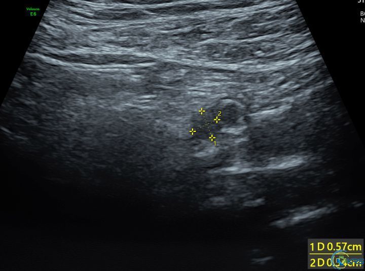
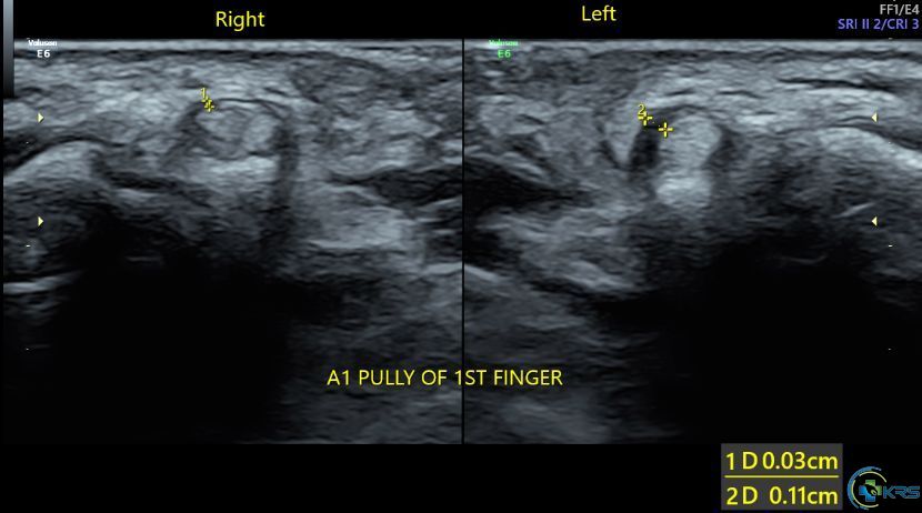
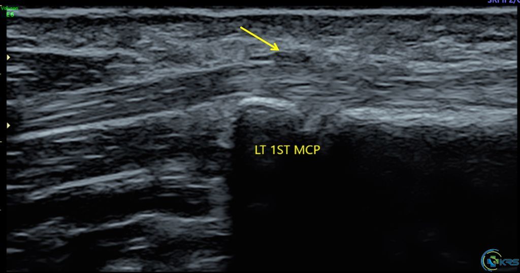
55 yr female presented with pain and stiffness of left thumb.
Ultrasound findings: