36 yr female presented with cyclical lower abdomen pain. History of previous LSCS.
Ultrasound findings:
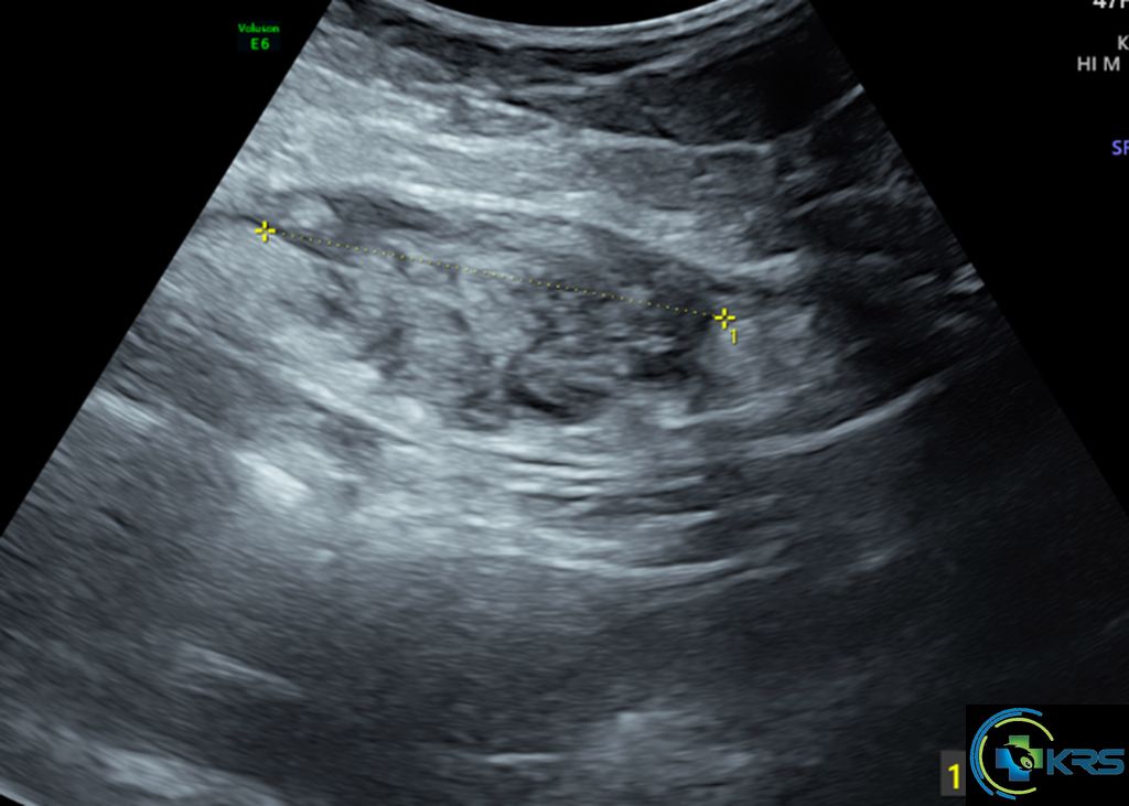
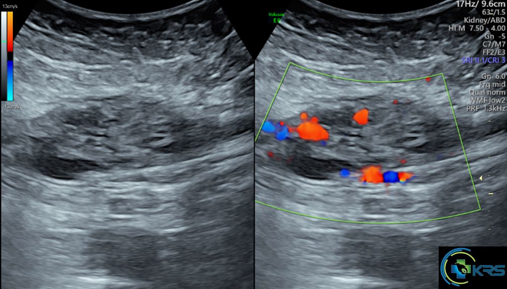
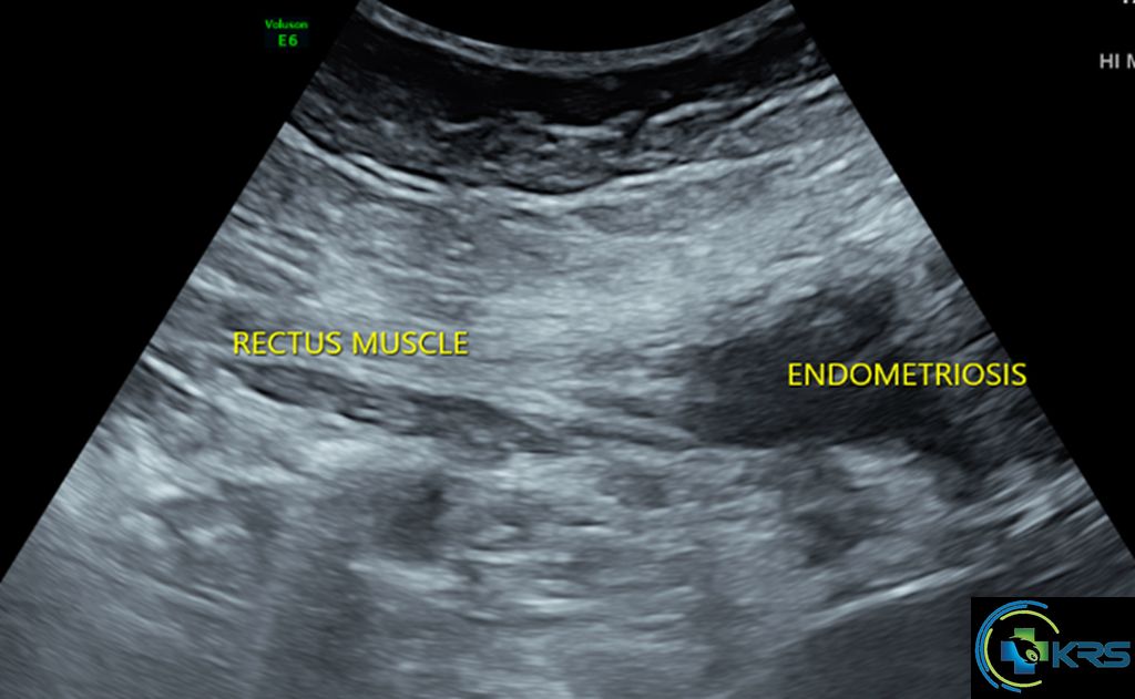
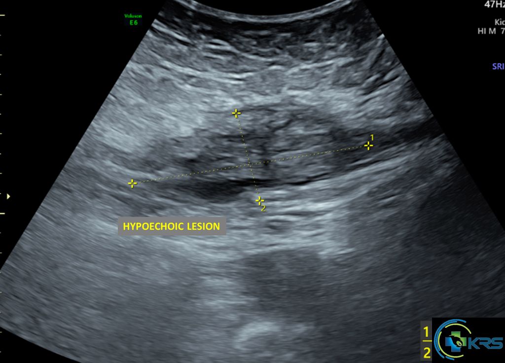
• Irregular heterogeneously hypoechoic lesion measuring 6 x 2.2 x 5 cm (SI x AP x T) with multiple small cystic areas and increased vascularity in muscle plane (involving the left rectus abdominis muscle) of anterior abdominal wall at lower abdomen – suggestive of scar endometriosis.
• The lesion is seen contained within the left rectus sheath.
• No evidence of extension into subcutaneous plane or intra-abdominal extension.
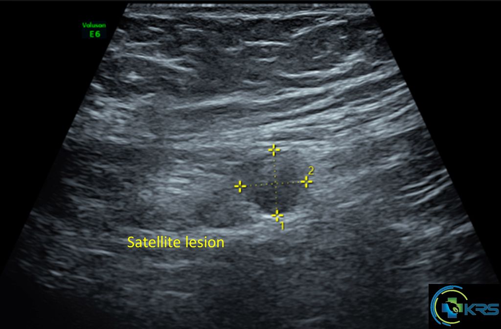
• Another small hypoechoic lesion measuring 1 x 1 cm in muscle plane 1.7 cm lateral to the above described lesion – satellite lesion.