3 yr boy presented with pain and swelling of left scrotum for 2 days.
Ultrasound findings:
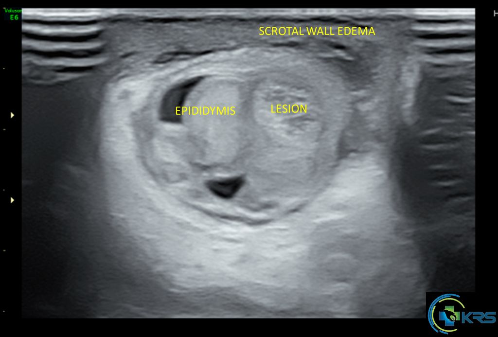
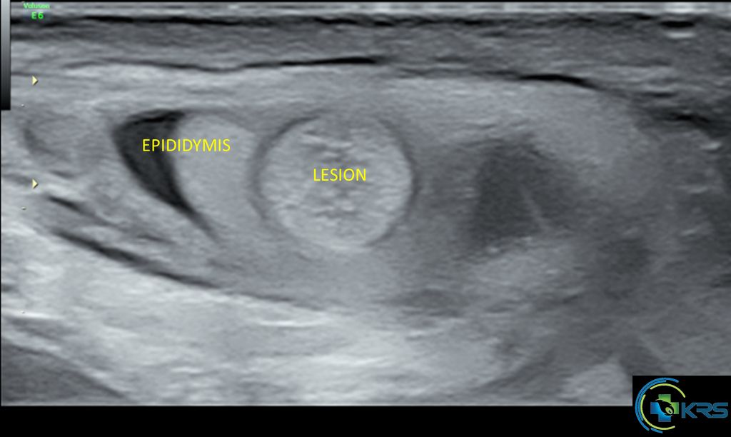
• A rounded well defined echogenic lesion measuring 7 x 6 mm noted in superior pole of left testis between head of epididymis and testis.
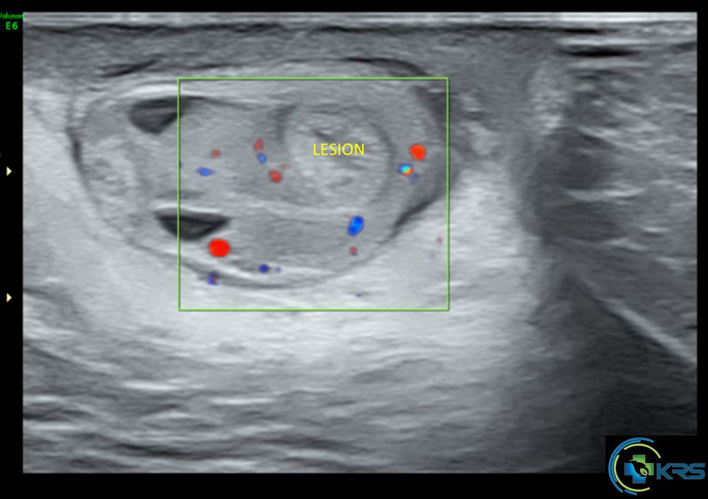
• Colour Doppler shows no internal vascularity within the lesion and normal vascularity of epididymis and testis.
• Minimal left hydrocele.
• Diffuse left scrotal wall edema.
• Feature suggestive of Torsion of left appendix testis.